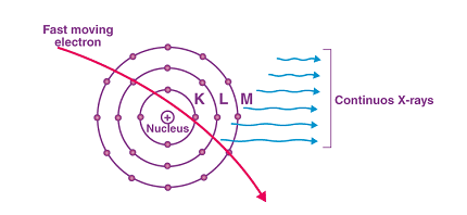
Continuous X-rays, also known as bremsstrahlung X-rays or simply “braking radiation,” are a type of X-ray radiation that is produced when a charged particle, such as an electron, is decelerated or slowed down by the electric field of an atomic nucleus or other charged particle. This process creates a broad spectrum of X-rays that range in energy from low to high, rather than the characteristic X-rays that are produced when an electron transitions from a higher energy level to a lower energy level in an atom.
Continuous X-rays are commonly used in medical imaging, such as in X-ray computed tomography (CT) scans, as well as in materials testing and other scientific applications. The energy of the X-rays produced depends on the energy of the incoming charged particle and the strength of the electric field it encounters. Therefore, higher-energy charged particles and stronger electric fields will produce higher-energy X-rays.
What is Continuous X-rays
Continuous X-rays, also known as bremsstrahlung X-rays or “braking radiation,” are a type of electromagnetic radiation that is produced when a charged particle, such as an electron, is decelerated or slowed down by the electric field of an atomic nucleus or other charged particle. This process creates a broad spectrum of X-rays that range in energy from low to high, rather than the characteristic X-rays that are produced when an electron transitions from a higher energy level to a lower energy level in an atom.
Continuous X-rays are commonly used in medical imaging, such as in X-ray computed tomography (CT) scans, as well as in materials testing and other scientific applications. The energy of the X-rays produced depends on the energy of the incoming charged particle and the strength of the electric field it encounters. Therefore, higher-energy charged particles and stronger electric fields will produce higher-energy X-rays.
When is Continuous X-rays
Continuous X-rays, also known as bremsstrahlung X-rays, can be produced in a variety of situations where charged particles are decelerated or slowed down by the electric field of an atomic nucleus or other charged particle. Some examples include:
- In medical imaging: Continuous X-rays are used in X-ray computed tomography (CT) scans to create detailed images of the internal structures of the body. In this application, a beam of X-rays is directed through the body, and the X-rays that pass through are detected and used to create an image.
- In materials testing: Continuous X-rays can be used to inspect the internal structure of materials, such as welds or castings, to detect flaws or defects that may compromise the structural integrity of the material.
- In scientific research: Continuous X-rays are used in a variety of scientific applications, including X-ray crystallography, which is used to determine the three-dimensional structure of molecules.
In all of these situations, the energy of the X-rays produced depends on the energy of the charged particles and the strength of the electric field they encounter. Higher-energy charged particles and stronger electric fields will produce higher-energy X-rays.
Where is Continuous X-rays
Continuous X-rays, also known as bremsstrahlung X-rays, can be found in a variety of settings where charged particles are decelerated or slowed down by the electric field of an atomic nucleus or other charged particle. Some common examples of where Continuous X-rays can be found include:
- Medical facilities: Continuous X-rays are used in medical imaging, such as in X-ray computed tomography (CT) scans, to create detailed images of the internal structures of the body.
- Industrial settings: Continuous X-rays can be used in industrial applications, such as in materials testing to inspect the internal structure of materials for defects or flaws.
- Scientific research facilities: Continuous X-rays are used in various scientific research applications, including X-ray crystallography to determine the three-dimensional structure of molecules.
- Airports and security checkpoints: Continuous X-rays are used in security checkpoints to scan luggage and other items for prohibited items.
- Research labs: Continuous X-rays can be found in research labs, where they are used in various experiments and scientific investigations.
In all of these settings, the energy of the X-rays produced depends on the energy of the charged particles and the strength of the electric field they encounter. Higher-energy charged particles and stronger electric fields will produce higher-energy X-rays.
How is Continuous X-rays
Continuous X-rays, also known as bremsstrahlung X-rays, are produced when a charged particle, such as an electron, is decelerated or slowed down by the electric field of an atomic nucleus or other charged particle. This deceleration causes the charged particle to lose energy, which is emitted in the form of X-rays.
The process of X-ray production through bremsstrahlung radiation can be described as follows:
- A charged particle, such as an electron, is accelerated towards an atomic nucleus or another charged particle.
- As the charged particle approaches the atomic nucleus or charged particle, it is deflected by the electric field and begins to slow down, losing kinetic energy.
- The lost kinetic energy is emitted as X-rays, with the energy of the X-rays depending on the energy of the incoming charged particle and the strength of the electric field it encounters.
- Because the deflection and deceleration of the charged particle is random, the resulting X-rays have a broad spectrum of energies, rather than a narrow set of energies like the characteristic X-rays produced by transitions between electron energy levels.
The resulting X-rays can be detected and used for a variety of applications, such as in medical imaging, materials testing, and scientific research.
Case Study on Continuous X-rays
One example of the use of continuous X-rays, or bremsstrahlung X-rays, is in medical imaging. X-ray computed tomography (CT) scans use a beam of X-rays, which includes both characteristic X-rays and continuous X-rays, to create detailed images of the internal structures of the body.
In a CT scan, a patient lies on a table that is moved through a ring-shaped machine that contains an X-ray source and a detector. The X-ray source emits a beam of X-rays, which pass through the patient’s body and are detected on the other side by the detector. The detector measures the intensity of the X-rays that pass through the patient and creates a two-dimensional image.
However, because the X-rays are emitted in all directions and pass through many different types of tissues with varying densities, the resulting image can be difficult to interpret. To create a more detailed image, the CT machine uses a technique called computed tomography, which combines many two-dimensional X-ray images taken from different angles to create a three-dimensional image of the body.
In computed tomography, the X-ray source rotates around the patient, emitting a series of X-ray beams at different angles. The detector measures the intensity of each beam, and a computer algorithm uses this information to create a three-dimensional image of the body. Because the X-ray beams include both characteristic X-rays and continuous X-rays, the resulting image contains information about the internal structures of the body that can be used to diagnose diseases and injuries.
Continuous X-rays are particularly useful in CT scans because they provide a broad spectrum of energies that can penetrate through many different types of tissues. This allows for a more detailed image of the internal structures of the body, including bones, soft tissues, and organs. However, because X-rays are ionizing radiation, which can potentially damage cells and increase the risk of cancer, CT scans should only be used when necessary and with appropriate radiation shielding and dosage control measures in place.
White paper on Continuous X-rays
Introduction
Continuous X-rays, also known as bremsstrahlung X-rays, are a type of X-ray radiation that is produced when a charged particle, such as an electron, is decelerated or slowed down by the electric field of an atomic nucleus or other charged particle. Continuous X-rays have a broad spectrum of energies, which makes them useful for a variety of applications, including medical imaging, materials testing, and scientific research. In this white paper, we will discuss the properties of continuous X-rays, their applications, and the safety considerations associated with their use.
Properties of Continuous X-rays
Continuous X-rays are produced when a charged particle is decelerated or slowed down by the electric field of an atomic nucleus or other charged particle. This process causes the charged particle to lose energy, which is emitted in the form of X-rays. The energy of the X-rays produced depends on the energy of the charged particle and the strength of the electric field it encounters. Higher-energy charged particles and stronger electric fields will produce higher-energy X-rays.
Unlike characteristic X-rays, which are produced when an electron transitions between energy levels in an atom, continuous X-rays have a broad spectrum of energies because the deceleration of the charged particle is random. This broad spectrum of energies makes continuous X-rays useful for a variety of applications, as they can penetrate through many different types of materials and tissues with varying densities.
Applications of Continuous X-rays
Continuous X-rays have a variety of applications, including:
- Medical imaging: Continuous X-rays are used in medical imaging, such as in X-ray computed tomography (CT) scans, to create detailed images of the internal structures of the body.
- Industrial settings: Continuous X-rays can be used in industrial applications, such as in materials testing to inspect the internal structure of materials for defects or flaws.
- Scientific research facilities: Continuous X-rays are used in various scientific research applications, including X-ray crystallography to determine the three-dimensional structure of molecules.
- Airport security checkpoints: Continuous X-rays are used in security checkpoints to scan luggage and other items for prohibited items.
- Research labs: Continuous X-rays can be found in research labs, where they are used in various experiments and scientific investigations.
Safety Considerations
Although continuous X-rays have many useful applications, they can also be potentially harmful. X-rays are ionizing radiation, which can damage cells and increase the risk of cancer. Therefore, it is important to use appropriate radiation shielding and dosage control measures when using continuous X-rays.
In medical settings, CT scans and other X-ray procedures should only be performed when necessary and with appropriate radiation shielding and dosage control measures in place. Patients should also be informed about the potential risks associated with X-ray radiation and given the opportunity to ask questions and make informed decisions about their care.
In industrial settings and research labs, appropriate safety measures should be taken to protect workers from exposure to X-ray radiation. This may include using radiation shielding, controlling the distance between workers and the X-ray source, and limiting the amount of time workers are exposed to X-ray radiation.
Conclusion
Continuous X-rays, or bremsstrahlung X-rays, are a type of X-ray radiation that is produced when a charged particle is decelerated or slowed down by the electric field of an atomic nucleus or other charged particle. Continuous X-rays have a broad spectrum of energies, which makes them useful for a variety of applications, including medical imaging, materials testing, and scientific research. However, because X-rays are ionizing radiation, which can potentially damage cells and increase the risk of cancer, it is important to use appropriate radiation shielding and dosage control measures when using continuous X-rays.