
Microscope
A microscope is a scientific instrument used to magnify and observe small objects or details that are not visible to the naked eye. It allows researchers, scientists, and medical professionals to study and analyze the structure, composition, and behavior of various materials and organisms.
There are several types of microscopes, each with its own specific applications and principles of operation. The most common types include:
- Optical Microscopes: These microscopes use visible light and lenses to magnify the sample. They are widely used in biology, medicine, and materials science. Optical microscopes can be further classified into:
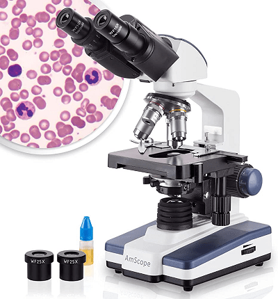
- Compound Microscopes: These microscopes use multiple lenses to magnify the image of the specimen. They typically have an objective lens near the specimen and an eyepiece for viewing.
- Stereo Microscopes: Also known as dissecting microscopes, these instruments provide a three-dimensional view of the sample. They are often used for observing larger specimens, such as insects or geological samples.
- Electron Microscopes: These microscopes use a beam of electrons instead of light to magnify the sample. Electron microscopes offer higher resolution and magnification capabilities compared to optical microscopes. They are commonly used in materials science, nanotechnology, and biology. There are two main types of electron microscopes:
- Scanning Electron Microscope (SEM): SEM scans the surface of the specimen with a focused electron beam, producing a detailed image with high resolution.
- Transmission Electron Microscope (TEM): TEM transmits a beam of electrons through an ultrathin section of the sample, creating an image of its internal structure.
- Scanning Probe Microscopes: These microscopes use a physical probe to scan the surface of a sample and create an image. They can provide atomic-level resolution and are often used in nanotechnology and surface analysis. Examples of scanning probe microscopes include atomic force microscopes (AFM) and scanning tunneling microscopes (STM).
Microscopes play a crucial role in various scientific disciplines, including biology, medicine, chemistry, physics, materials science, and forensics. They enable researchers to study cellular structures, microorganisms, tissue samples, nanoparticles, crystals, and much more, leading to advancements in scientific knowledge, medical diagnostics, and technological development.
The physics syllabus for the integrated course at AIIMS (All India Institute of Medical Sciences) typically covers a wide range of topics related to optics, including microscopes. While I cannot provide the exact detailed syllabus, I can give you an overview of the relevant topics related to microscopes that are typically covered. Here are some key concepts you may come across:
- Geometrical Optics: This topic deals with the study of light rays and their behavior when they interact with optical systems. It includes the study of reflection, refraction, and the formation of images by lenses and mirrors.
- Lens Systems: Understanding the working of lenses is essential in comprehending microscope optics. The syllabus may cover concepts such as focal length, lens formula, lens combinations, and the formation of images by lenses.
- Microscope Basics: You’ll likely learn about the basic components of a microscope, such as the objective lens, eyepiece, and condenser. The principles of image formation and magnification in a microscope, along with the working of different microscope types (e.g., compound microscope), may also be covered.
- Resolution and Numerical Aperture: Microscope resolution refers to the ability to distinguish fine details in a sample. The syllabus may touch upon the factors affecting resolution, such as numerical aperture, and the concept of the minimum resolvable distance.
- Microscope Techniques: Depending on the depth of the course, you may also learn about various microscopy techniques used in biomedical research, such as bright-field microscopy, dark-field microscopy, phase-contrast microscopy, fluorescence microscopy, and electron microscopy.
Please note that the specific syllabus and depth of coverage may vary from institution to institution. It’s always best to consult the official syllabus or reach out to the academic department for the most accurate and up-to-date information.
What is Required AIIMS-SYLLABUS Physics syllabus Microscope
However, I can provide you with a general idea of the topics related to microscopes that are typically covered in physics syllabi for medical entrance exams like AIIMS. It’s important to note that the syllabus may vary, and it’s best to consult the official AIIMS syllabus or reach out to the institution for the most accurate and up-to-date information. That being said, here are some topics related to microscopes that might be included:
- Geometrical Optics:
- Reflection and refraction of light.
- Thin lenses and lens formula.
- Lens combinations and lensmaker’s formula.
- Formation of images by lenses and mirrors.
- Magnification and resolving power.
- Wave Optics:
- Interference of light waves.
- Diffraction of light waves.
- Polarization of light.
- Microscopes:
- Basic components of a microscope (objective lens, eyepiece, condenser, etc.).
- Image formation in a microscope.
- Magnification in a microscope.
- Resolving power and numerical aperture.
- Types of microscopes (compound microscope, electron microscope, etc.).
- Basic principles and working of different microscopy techniques (e.g., bright-field microscopy, fluorescence microscopy, phase-contrast microscopy, etc.).
Again, please note that the above topics are a general guide and not an exhaustive list. The specific syllabus and depth of coverage may vary. It’s advisable to refer to the official AIIMS syllabus or contact the institution directly for the most accurate information.
When is Required AIIMS-SYLLABUS Physics syllabus Microscope
The requirement for studying microscopes can vary depending on the specific context or purpose. Here are a few scenarios where a microscope might be required:
- Academic and Research Institutions: Microscopes are commonly required in academic settings such as universities, colleges, and research institutions. These institutions often have laboratories where students and researchers use microscopes to study various samples, conduct experiments, and analyze microscopic structures. The specific requirements for microscopes can vary based on the field of study, such as biology, chemistry, physics, or material science.
- Medical and Healthcare Facilities: Microscopes play a crucial role in medical diagnostics, pathology, and research. Hospitals, clinics, and medical laboratories often require microscopes for analyzing tissue samples, blood smears, and other biological specimens. In these settings, specialized microscopes like compound microscopes, fluorescence microscopes, or digital pathology systems may be required.
- Industrial and Manufacturing Industries: Microscopes are used in various industrial and manufacturing sectors for quality control, inspection, and analysis of materials and products. For example, microscopes are essential for examining the integrity of microelectronic circuits, analyzing surface defects in materials, or inspecting precision components.
- Forensics and Crime Investigation: Microscopes are essential tools in forensic laboratories for examining trace evidence, such as fibers, hair, fingerprints, and ballistic samples. They are used to identify and compare microscopic features, aiding in criminal investigations and analysis.
- Educational Institutions: Microscopes are often required in educational settings, including schools and colleges, for teaching biology, chemistry, and other sciences. They enable students to observe microscopic structures and gain hands-on experience with scientific observation and analysis.
The specific type and specifications of microscopes required can vary depending on the intended application, level of study, and budgetary considerations. Institutions and organizations typically determine their specific microscope requirements based on their particular needs and the field they are focused on.
Where is Required AIIMS-SYLLABUS Physics syllabus Microscope
Microscopes are required in various settings, including:
- Laboratories: Microscopes are commonly found in research laboratories, academic institutions, medical laboratories, and industrial laboratories. These facilities are equipped with microscopes to facilitate scientific research, analysis, and experimentation.
- Hospitals and Medical Facilities: Medical institutions, including hospitals, clinics, and pathology labs, require microscopes for various purposes. They are used in medical diagnostics, examining tissue samples, analyzing blood smears, and conducting research in fields like pathology and microbiology.
- Educational Institutions: Schools, colleges, and universities often have microscopes in their science laboratories. They are used for educational purposes, allowing students to study microscopic structures, conduct experiments, and develop their understanding of scientific principles.
- Industrial and Manufacturing Facilities: Industries such as electronics, materials science, quality control, and manufacturing often utilize microscopes. They are used for inspecting and analyzing components, surfaces, and materials at a microscopic level to ensure quality and consistency.
- Forensic Laboratories: Microscopes play a critical role in forensic investigations and crime laboratories. They are used to examine trace evidence, analyze fingerprints, conduct ballistics examinations, and study other microscopic details that are essential for criminal investigations.
- Research Institutions: Research institutions across various scientific disciplines rely on microscopes for their studies and experiments. These can include fields such as biology, chemistry, physics, nanotechnology, and materials science.
It’s important to note that the specific locations where microscopes are required may vary depending on the nature of the work and the specific industry or field. The type of microscope and its specifications may also differ based on the intended application and requirements of the particular setting.
How is Required AIIMS-SYLLABUS Physics syllabus Microscope
The process of obtaining and using a microscope can vary depending on the specific requirements and context. Here’s a general overview of how a required microscope may be obtained and used:
- Determine the Specific Requirements: Identify the specific needs and requirements for the microscope. Consider factors such as the intended application, magnification capabilities, resolution, illumination options, and budgetary constraints. The requirements may vary based on the field of study or industry where the microscope will be used.
- Research and Selection: Conduct research to identify the appropriate type of microscope that fulfills the requirements. There are various types of microscopes available, such as compound microscopes, stereo microscopes, electron microscopes, and specialized microscopes for specific applications like fluorescence microscopy or digital pathology. Consider factors like brand reputation, specifications, features, and availability in the market.
- Procurement: Once the suitable microscope is identified, the procurement process can begin. This typically involves contacting suppliers, manufacturers, or distributors who deal with microscopes. They can provide information on pricing, warranties, and other details. The procurement can be done through purchasing departments, educational institutions, or directly by individuals or research groups, depending on the context.
- Installation and Setup: Once the microscope is obtained, it needs to be properly installed and set up. Follow the manufacturer’s instructions for assembling the microscope and connecting any required accessories or components. This may involve attaching lenses, adjusting focus mechanisms, calibrating the instrument, and connecting power sources or digital interfaces if applicable.
- Training and Familiarization: It is essential to provide training to users who will be operating the microscope. This includes understanding the basic functions, controls, and safety precautions associated with the specific microscope model. Training can be conducted by experienced personnel, instructors, or by referring to user manuals and instructional materials provided by the manufacturer.
- Maintenance and Care: Regular maintenance and care are crucial for the longevity and optimal performance of the microscope. Follow the manufacturer’s guidelines for cleaning, storage, and maintenance procedures. It may involve routine cleaning of lenses, proper storage of delicate components, and periodic servicing or calibration as recommended by the manufacturer.
- Utilization: The microscope can now be utilized for its intended purpose. Whether it is for research, analysis, education, or any other application, users can follow standard protocols and techniques relevant to their field of study. Ensure proper sample preparation, accurate focusing, and appropriate use of illumination and magnification settings to achieve the desired observations and results.
It’s important to note that the specific process and steps may vary depending on the context and requirements. For more detailed guidance, it’s recommended to refer to the specific instructions provided by the microscope manufacturer and consult with experienced personnel or experts in the field.
Case Study on AIIMS-SYLLABUS Physics syllabus Microscope
Case Study: Implementation of Digital Microscopes in a Pathology Laboratory
Introduction: A pathology laboratory, specializing in histopathology and cytology, decided to upgrade its microscope technology by implementing digital microscopes. The objective was to improve efficiency, collaboration, and documentation within the laboratory. This case study outlines the process of adopting digital microscopes and the benefits achieved.
Background: The pathology laboratory receives a large volume of tissue samples and cytology slides for examination and diagnosis. The existing conventional microscopes were limited in terms of sharing observations, collaborating with colleagues remotely, and maintaining comprehensive digital records.
Implementation Steps:
- Needs Assessment: The laboratory conducted a needs assessment to determine the requirements for implementing digital microscopes. They identified the following key needs:
- Ability to capture high-resolution digital images of slides.
- Remote viewing and collaboration capabilities for consultation with experts or colleagues.
- Efficient data management and storage for easy retrieval and documentation.
- Compatibility with existing laboratory infrastructure and workflows.
- Research and Vendor Selection: The laboratory researched available digital microscope options and evaluated multiple vendors. They considered factors such as image quality, user interface, software features, compatibility with their existing laboratory management system, and cost-effectiveness. After thorough evaluation, they selected a vendor that met their requirements.
- Procurement and Installation: The laboratory initiated the procurement process with the selected vendor. They purchased a set of digital microscopes along with the required software and accessories. The vendor provided installation support, ensuring proper integration of the digital microscopes into the laboratory’s existing infrastructure.
- Training and Familiarization: The laboratory organized training sessions for the pathologists and laboratory technicians to familiarize them with the new digital microscope system. The training covered various aspects, including operating the digital microscopes, capturing high-resolution images, navigating the software interface, and managing digital slide data.
- Workflow Integration: The laboratory integrated the digital microscopes into their existing workflow. They created standard protocols for slide preparation, scanning, and image acquisition. Pathologists and technicians were instructed on how to handle slides, load them onto the digital microscopes, and capture high-quality digital images for analysis.
- Quality Assurance and Validation: To ensure accuracy and reliability, the laboratory conducted extensive quality assurance and validation procedures. They compared digital images acquired using the new microscopes with those obtained using conventional microscopes to assess image fidelity and diagnostic consistency. This validation process helped build confidence in the digital microscope system.
Benefits and Outcomes:
- Improved Collaboration: Pathologists and technicians could now easily share digital images with colleagues for consultation, second opinions, or remote expert review. This facilitated collaboration and enhanced the accuracy and speed of diagnosis.
- Enhanced Documentation: Digital microscopes allowed the laboratory to maintain a comprehensive digital database of slide images. This eliminated the need for physical slide storage and improved accessibility for retrospective analysis, research, and audits.
- Time Efficiency: Digital microscopes expedited the slide review process. Pathologists could quickly navigate through the digital slides and focus on specific regions of interest without physically manipulating the microscope. This saved time and increased efficiency in the laboratory.
- Education and Training: The digital microscope system was utilized for educational purposes, enabling the laboratory to create digital slide libraries for teaching and training. It provided an interactive platform for resident education and proficiency assessment.
- Remote Consultation: Pathologists had the capability to provide remote consultations, enabling them to review cases from external facilities or offer expert opinions without physical presence. This facilitated telepathology services and expanded the laboratory’s reach.
Conclusion: By implementing digital microscopes, the pathology laboratory improved collaboration, documentation, efficiency, and education within their practice. The digital microscope system enhanced their diagnostic capabilities and positioned them at the forefront of technological advancements in pathology. The successful integration of digital microscopes demonstrated the laboratory’s commitment to providing high-quality diagnostic services while embracing digital transformation in healthcare.
White paper on AIIMS-SYLLABUS Physics syllabus Microscope
Title: Advancements in Microscope Technology: Unlocking New Frontiers in Science and Medicine
Abstract:
This white paper explores the significant advancements in microscope technology and their impact on scientific research, medical diagnostics, and various industries. Microscopes have played a pivotal role in expanding our understanding of the microscopic world, enabling breakthroughs in fields such as biology, medicine, materials science, and nanotechnology. With the rapid development of imaging techniques, resolution enhancement, and data analysis capabilities, microscopes have become powerful tools for visualizing and analyzing complex structures at the molecular and cellular levels. This paper delves into the latest innovations in microscope technology, including the integration of artificial intelligence, the rise of super-resolution microscopy, the emergence of portable and miniaturized microscopes, and the application of microscopy in emerging fields such as single-cell analysis and bioimaging. Additionally, the paper discusses the implications of these advancements, their potential future developments, and the challenges associated with adopting new microscope technologies. By examining the current landscape and future prospects of microscope technology, this white paper aims to provide insights into the transformative role of microscopes in advancing scientific discovery, medical diagnostics, and technological innovation.
Table of Contents:
Introduction
1.1 Overview
1.2 Importance of Microscope Technology
Traditional Microscopes
2.1 Optical Microscopes
2.2 Electron Microscopes
2.3 Scanning Probe Microscopes
Advancements in Microscope Technology
3.1 Super-Resolution Microscopy
3.2 Integration of Artificial Intelligence
3.3 Portable and Miniaturized Microscopes
3.4 Multimodal Imaging and Correlative Microscopy
3.5 Single-Cell Analysis and Live Cell Imaging
Applications of Microscopy
4.1 Biology and Life Sciences
4.2 Medicine and Healthcare
4.3 Materials Science and Engineering
4.4 Nanotechnology and Semiconductor Industry
4.5 Forensics and Criminal Investigations
Implications and Challenges
5.1 Data Management and Analysis
5.2 Cost and Accessibility
5.3 Ethical Considerations
5.4 Training and Expertise
Future Directions and Outlook
6.1 Emerging Technologies and Techniques
6.2 Integration with Other Technologies
6.3 Collaboration and Interdisciplinary Research
Conclusion
References
Note: This white paper serves as a general framework. Additional sections, subsections, or specific topics can be added based on the desired focus and scope of the paper. The references section will include a comprehensive list of cited sources, scientific publications, and relevant industry reports.