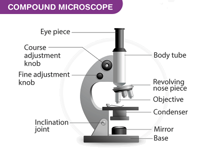
Microscope

A microscope is an optical instrument used to observe and magnify tiny objects that are otherwise difficult to see with the naked eye. It consists of several components that work together to produce a magnified image of the specimen being observed. The two main types of microscopes are optical microscopes and electron microscopes.
Optical Microscopes:
- Light Microscopes: They use visible light to illuminate the specimen. Light passes through the specimen and a series of lenses, including an objective lens and an eyepiece, to produce a magnified image. Different techniques like bright-field, phase contrast, dark-field, and fluorescence microscopy can be used to enhance contrast and visualize specific components of the specimen.
Electron Microscopes:
- Transmission Electron Microscope (TEM): It uses a beam of electrons to pass through the specimen. The electrons interact with the specimen, forming an image that is projected onto a fluorescent screen or a digital camera. TEM provides higher magnification and resolution compared to light microscopes and is used to study the internal structure of cells and materials.
- Scanning Electron Microscope (SEM): It scans a focused beam of electrons across the surface of the specimen. As the electrons interact with the specimen, secondary electrons are emitted and captured to form an image. SEM provides a detailed three-dimensional surface view of the specimen.
Components of a Microscope:
- Objective Lens: It collects and magnifies light or electrons from the specimen.
- Eyepiece: It further magnifies the image produced by the objective lens and allows the viewer to observe the specimen.
- Condenser: It focuses and directs light onto the specimen in an optical microscope or controls the electron beam in an electron microscope.
- Stage: It holds the specimen in place for observation.
- Illumination Source: In optical microscopes, it provides the light necessary to illuminate the specimen. It can be a light bulb, an LED, or a laser, depending on the microscope type.
Applications of Microscopes: Microscopes have extensive applications in various fields, including:
- Biology: Studying cells, tissues, and microorganisms.
- Medicine: Diagnosis and analysis of diseases.
- Material Science: Analyzing the structure and properties of materials.
- Forensics: Examining evidence in criminal investigations.
- Nanotechnology: Characterizing nanoscale structures and devices.
- Quality Control: Inspecting and analyzing products for defects or abnormalities.
Microscopes play a crucial role in advancing scientific research, medical diagnostics, and technological development by enabling the visualization and study of microscopic objects and phenomena.
The physics syllabus for the AIIMS entrance exam typically includes the topic of the microscope. Here is a concise overview of the microscope topic:
Microscope:
- Introduction to Microscopes: Principles and types of microscopes (light microscope, electron microscope, etc.).
- Optical Microscopes: Ray optics, lens equation, magnification, resolving power, numerical aperture, and working principles of compound microscopes.
- Electron Microscopes: Principles and working of transmission electron microscope (TEM) and scanning electron microscope (SEM).
- Microscope Components: Detailed study of microscope components like objective lens, eyepiece, condenser, stage, and their functions.
- Image Formation: Formation of real and virtual images by microscopes, ray diagrams, and calculations related to magnification and image distance.
- Abbe’s Theory of Image Formation: Abbe’s resolution criterion, resolving power, and its dependence on wavelength and numerical aperture.
- Depth of Field and Depth of Focus: Definitions and calculations related to depth of field and depth of focus in microscopy.
- Techniques and Applications: Techniques like phase contrast microscopy, dark-field microscopy, fluorescence microscopy, and their applications in various fields.
- Scanning Probe Microscopy: Basics of atomic force microscopy (AFM) and scanning tunneling microscopy (STM).
- Recent Advancements: Brief introduction to advanced microscopy techniques such as confocal microscopy, super-resolution microscopy, and electron microscopy with cryo-EM.
Note: This is a general overview and the actual AIIMS syllabus may vary slightly. It is always recommended to refer to the official syllabus provided by AIIMS or the exam conducting authority for the most accurate and up-to-date information.
What is Required Physics syllabus Microscope
The required physics syllabus for studying microscopes typically includes the following topics:
- Optics:
- Geometrical optics: Ray optics, lens equation, magnification, and image formation by lenses.
- Wave optics: Interference, diffraction, and polarization of light.
- Microscope Basics:
- Types of microscopes: Optical microscopes (including compound microscopes) and electron microscopes.
- Components of microscopes: Objective lens, eyepiece, condenser, stage, and illumination system.
- Optical Microscopes:
- Working principle of optical microscopes.
- Magnification and resolving power.
- Numerical aperture and its importance.
- Bright-field microscopy: Basic concepts and image formation.
- Phase contrast microscopy: Principles and applications.
- Dark-field microscopy: Principles and applications.
- Fluorescence microscopy: Basic principles and applications.
- Electron Microscopes:
- Transmission Electron Microscope (TEM): Working principle and applications.
- Scanning Electron Microscope (SEM): Working principle and applications.
- Advanced Microscopy Techniques:
- Confocal microscopy: Principles and applications.
- Super-resolution microscopy: Principles and applications.
- Atomic Force Microscopy (AFM): Basics and applications.
- Scanning Tunneling Microscopy (STM): Basics and applications.
It’s important to note that the specific syllabus may vary depending on the educational institution or the exam you are preparing for. It is always recommended to refer to the official syllabus provided by the relevant authority or institution for the most accurate and up-to-date information.
When is Required Physics syllabus Microscope
The required physics syllabus for studying microscopes is typically covered in physics courses or curricula at the college or university level. The specific timing of when this topic is included in the syllabus can vary depending on the educational institution and the structure of the physics program.
In general, the study of microscopes and related topics is typically introduced after covering fundamental principles of optics, such as geometrical optics and wave optics. This often occurs in the later part of an introductory physics course or in a specialized course focused on optics or experimental techniques.
For students pursuing a specific field of study related to microscopy, such as biomedical sciences or materials science, the topic of microscopes may be covered in more detail in specialized courses within their respective disciplines.
It’s important to check the specific curriculum or syllabus of the physics course or program you are enrolled in or planning to pursue to determine the exact timing and depth of coverage of the microscope topic.
Where is Required Physics syllabus Microscope
The required physics syllabus that includes the topic of microscopes can be found in various educational settings. Here are some common places where you can find the physics syllabus that covers microscopes:
- Academic Institutions: Physics syllabi, including the microscope topic, are typically provided by academic institutions such as colleges, universities, and technical schools. These institutions usually have physics departments or programs that outline the specific topics covered in their physics courses. You can find the syllabus either on their official websites or by contacting the respective physics department.
- Entrance Exam Preparation Materials: If you are preparing for a specific entrance exam that includes physics, such as the AIIMS entrance exam mentioned earlier, the examining authority or the organization responsible for conducting the exam may provide a detailed syllabus. These syllabi often specify the topics to be studied, including the microscope topic.
- Textbooks and Course Materials: Physics textbooks and course materials can provide a comprehensive overview of the required syllabus. Look for textbooks that cover optics, modern physics, or specialized texts on microscopy and imaging techniques. These resources often outline the relevant topics related to microscopes and provide detailed explanations and examples.
- Online Educational Platforms: Various online educational platforms offer physics courses or learning materials that cover a wide range of topics, including microscopes. These platforms may provide detailed syllabi or course outlines that specify the topics covered. Examples of such platforms include Coursera, Khan Academy, edX, and Udemy.
When searching for the required physics syllabus on microscopes, it’s important to consider the specific context, such as the educational institution, entrance exam, or learning platform you are associated with. Checking official sources and consulting with instructors or academic advisors can help you access the most accurate and up-to-date information on the required syllabus.
How is Required Physics syllabus Microscope
The required physics syllabus for studying microscopes is typically structured to provide a comprehensive understanding of the principles, operation, and applications of microscopes. Here is a general outline of how the microscope topic may be covered in the physics syllabus:
- Introduction to Optics:
- Review of basic optics principles, including ray optics and wave optics.
- Geometrical optics: Lens equation, magnification, and image formation by lenses.
- Wave optics: Interference, diffraction, and polarization of light.
- Optical Microscopes:
- Overview of different types of microscopes: Optical microscopes (including compound microscopes) and electron microscopes.
- Study of microscope components: Objective lens, eyepiece, condenser, stage, and illumination system.
- Working principle of optical microscopes: Light path, specimen illumination, and image formation.
- Magnification and resolving power: Definitions, calculations, and limitations.
- Techniques and applications:
- Bright-field microscopy: Basic principles and image formation.
- Phase contrast microscopy: Principles, phase plate, and applications.
- Dark-field microscopy: Principles and applications.
- Fluorescence microscopy: Principles, fluorophores, and applications.
- Electron Microscopes:
- Introduction to electron microscopy: Transmission Electron Microscope (TEM) and Scanning Electron Microscope (SEM).
- Working principles of TEM and SEM: Electron beam formation, interaction with the specimen, and image formation.
- Applications and advantages of electron microscopy over optical microscopy.
- Advanced Microscopy Techniques:
- Confocal microscopy: Principles, laser scanning, and applications.
- Super-resolution microscopy: Principles (e.g., STED, PALM, STORM), resolution enhancement, and applications.
- Atomic Force Microscopy (AFM): Basics, tip-sample interaction, and applications.
- Scanning Tunneling Microscopy (STM): Basic principles, tunneling current, and applications.
The syllabus may also include practical aspects such as microscope alignment, sample preparation, and image analysis techniques.
It’s important to note that the specific organization and order of topics within the syllabus may vary depending on the educational institution, course structure, and the level of study (e.g., undergraduate or graduate level). Always refer to the official syllabus provided by your educational institution or the specific exam you are preparing for to obtain the most accurate and detailed information on the required physics syllabus for microscopes.
Case Study on Physics syllabus Microscope
Case Study: Application of Microscopes in Biomedical Research
Introduction: Microscopes play a vital role in biomedical research, enabling scientists and researchers to study cellular structures, organisms, and diseases at a microscopic level. This case study explores the application of microscopes in a specific biomedical research project involving the study of cancer cells and their response to drug treatment.
Objective: The objective of the study is to investigate the cellular and molecular changes in cancer cells when subjected to a novel drug treatment. The researchers aim to understand the drug’s mechanism of action, its effect on cell viability, and potential cellular alterations induced by the treatment.
Methods:
- Cell Culture: Cancer cells of interest are cultured in a controlled laboratory environment.
- Drug Treatment: The cancer cells are exposed to the novel drug at different concentrations and durations.
- Viability Assay: The viability of cells under different drug conditions is assessed using a fluorescence-based assay. Live cells are labeled with a fluorescent dye, and a fluorescence microscope is used to quantify the number of live and dead cells.
- Morphological Analysis: The treated cells are fixed, stained, and examined under an optical microscope. Morphological changes such as cell shape, size, and cellular structures are observed and documented.
- Immunofluorescence Staining: Specific cellular components or proteins of interest are labeled with fluorescent markers. Microscopy techniques like confocal microscopy or fluorescence microscopy are employed to visualize and quantify changes in protein expression or localization.
- Time-lapse Imaging: The drug-treated cells are imaged at regular intervals using time-lapse microscopy. This allows researchers to observe dynamic cellular processes, such as cell division, migration, or apoptosis, over time.
- Electron Microscopy: For a more detailed analysis of cellular structures, transmission electron microscopy (TEM) is employed to examine ultrastructural changes induced by the drug treatment.
- Image Analysis: Advanced image analysis software is used to quantify and analyze the collected microscopic images. Parameters such as cell size, shape, protein expression levels, and subcellular localization are quantified and statistically analyzed.
Results and Findings: The microscopy-based analysis of the drug-treated cancer cells reveals several significant findings:
- The drug treatment leads to a decrease in cell viability in a dose-dependent manner.
- Morphological analysis demonstrates alterations in cell shape, membrane integrity, and cellular organelles.
- Immunofluorescence staining reveals changes in the expression and localization of specific proteins associated with cell proliferation, apoptosis, or drug resistance.
- Time-lapse imaging shows delayed cell division, decreased cell motility, and increased apoptotic events.
- Electron microscopy reveals ultrastructural changes in the treated cells, such as mitochondrial abnormalities and disrupted endoplasmic reticulum.
Conclusion: This case study demonstrates the essential role of microscopes in biomedical research. The application of various microscopy techniques, including optical microscopy, fluorescence microscopy, and electron microscopy, allows researchers to visualize and analyze cellular and molecular changes in cancer cells undergoing drug treatment. The microscopic observations and quantitative analysis provide valuable insights into the drug’s mechanism of action, cellular responses, and potential therapeutic implications. Such studies contribute to the development of targeted and effective cancer treatments.
Note: This case study provides a general example of how microscopes are applied in biomedical research. The specific details, experimental design, and findings can vary based on the research project, objectives, and techniques employed.
White paper on Physics syllabus Microscope
Title: Advancements in Microscopy: Unlocking the Invisible World
Abstract: Microscopy has been an indispensable tool in scientific research, enabling us to explore and understand the intricate structures and phenomena of the microscopic world. This white paper explores the advancements in microscopy technologies, their applications across various fields, and their potential impact on scientific discoveries and technological advancements.
- Introduction:
- Importance of microscopy in scientific research and technological development.
- Overview of the historical evolution of microscopes.
- Optical Microscopy:
- Principles of optical microscopy, including bright-field, phase contrast, and fluorescence microscopy.
- Recent advancements in optical microscopy techniques, such as confocal microscopy, super-resolution microscopy, and light sheet microscopy.
- Applications in biology, medicine, materials science, and nanotechnology.
- Electron Microscopy:
- Introduction to electron microscopy, including scanning electron microscopy (SEM) and transmission electron microscopy (TEM).
- High-resolution imaging and analysis of biological specimens, cells, tissues, and materials at the nanoscale.
- Cryo-electron microscopy (cryo-EM) and its impact on structural biology and drug discovery.
- Scanning Probe Microscopy:
- Atomic force microscopy (AFM) and scanning tunneling microscopy (STM) principles.
- Imaging and manipulation of surfaces and materials at the atomic and molecular level.
- Applications in nanoscience, surface analysis, and materials characterization.
- Correlative Microscopy:
- Integration of multiple microscopy techniques for complementary analysis.
- Combining optical microscopy, electron microscopy, and spectroscopy for comprehensive sample characterization.
- Advantages and challenges of correlative microscopy.
- Live Cell Imaging:
- Real-time imaging of cellular processes and dynamics.
- Fluorescence-based techniques, time-lapse microscopy, and single-molecule imaging.
- Applications in cell biology, neuroscience, and drug discovery.
- Data Analysis and Image Processing:
- Importance of advanced image analysis algorithms and software.
- Automation, quantification, and extraction of meaningful information from microscopy data.
- Machine learning and artificial intelligence in microscopy analysis.
- Emerging Trends and Future Directions:
- In situ and in vivo imaging techniques.
- Integration of microscopy with other analytical techniques, such as spectroscopy and mass spectrometry.
- Lab-on-a-chip and miniaturized microscopy devices.
- Advancements in sample preparation techniques and imaging modalities.
- Conclusion:
- Summary of the advancements in microscopy and their impact on scientific research and technological innovations.
- Future prospects and challenges in the field of microscopy.
This white paper provides an overview of the advancements in microscopy technologies, their applications, and their potential to unlock new insights into the microscopic world. By continuously pushing the boundaries of resolution, sensitivity, and imaging capabilities, microscopy plays a pivotal role in driving scientific discoveries and technological advancements in various disciplines.