
X-rays are a form of electromagnetic radiation with wavelengths shorter than those of visible light. They were discovered by Wilhelm Conrad Roentgen in 1895 and have since become an important tool in medicine, industry, and scientific research.
In medicine, X-rays are used to create images of the inside of the body. This allows doctors to diagnose and treat a wide range of medical conditions, such as bone fractures, tumors, and dental problems. However, repeated exposure to X-rays can be harmful and increase the risk of cancer.
In industry, X-rays are used to inspect objects for flaws or defects. For example, they can be used to check the quality of welds in pipelines or the integrity of airplane wings. X-rays are also used in scientific research to study the structure of materials and the behavior of atoms and molecules.
Overall, X-rays are a powerful tool with many applications, but their use should be carefully controlled to minimize the risk of harm.
What is X-Rays
X-rays are a type of high-energy electromagnetic radiation with wavelengths shorter than those of visible light. They were discovered by Wilhelm Conrad Roentgen in 1895 and have since become an important tool in many fields, including medicine, industry, and scientific research.
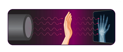
X-rays are produced when high-energy electrons are accelerated and collide with a metal target. The collision causes the electrons to release energy in the form of X-rays. These X-rays can penetrate solid objects, and their ability to pass through different materials varies depending on the material’s density and thickness.
In medicine, X-rays are used to create images of the inside of the body, allowing doctors to diagnose and treat a wide range of conditions. In industry, X-rays are used to inspect objects for flaws or defects, such as cracks or imperfections in metal parts. In scientific research, X-rays are used to study the structure of materials, the behavior of atoms and molecules, and many other phenomena.
While X-rays are useful in many applications, they can also be harmful. Repeated exposure to X-rays can increase the risk of cancer, and precautions must be taken to ensure that X-ray doses are kept as low as possible.
When is X-Rays
X-rays can be used in a wide range of applications, including medical imaging, industrial testing, and scientific research.
In medicine, X-rays are commonly used to create images of bones and other internal structures in the body. This can help doctors diagnose and treat a variety of medical conditions, including broken bones, dental problems, and tumors.
In industry, X-rays are used to inspect the quality of materials and products, such as welds in pipelines, electronic components, and aircraft parts. X-rays can also be used to identify defects or weaknesses in metal structures.
In scientific research, X-rays can be used to study the structure of materials at the atomic level. This can help scientists understand the properties of materials and develop new technologies.
Overall, X-rays can be used whenever it is necessary to see inside an object or to study its properties at a very small scale. However, it is important to use X-rays carefully and follow appropriate safety protocols to minimize the risk of harm from radiation exposure.
Where is X-Rays
X-rays can be found and used in many different locations and settings, depending on the specific application.
In medicine, X-rays are typically performed in a hospital, clinic, or medical imaging center. These facilities have specialized equipment that can produce and detect X-rays, and trained professionals who can interpret the images to help diagnose and treat medical conditions.
In industry, X-rays may be used in manufacturing plants or testing facilities to inspect products and materials. Specialized X-ray machines or instruments may be used for these applications, and trained personnel may be required to operate them safely.
In scientific research, X-rays are often used at specialized research facilities, such as synchrotron radiation sources or X-ray crystallography laboratories. These facilities have highly specialized equipment that can produce and manipulate X-rays at very high energies and intensities, allowing scientists to study the structure and behavior of materials and molecules at a very small scale.
Overall, X-rays can be found in a wide range of locations and settings, depending on the application and the specific equipment or technology used.
How is X-Rays
X-rays are typically produced using a specialized machine called an X-ray generator. The X-ray generator includes a vacuum tube that contains a cathode and an anode.
When an electric current is applied to the cathode, it produces a stream of high-energy electrons. The electrons are then focused onto a small target on the anode, which is typically made of a heavy metal such as tungsten. When the electrons collide with the anode, they produce X-rays.
The X-rays produced by the generator are then directed through the object being studied, such as a patient’s body or a metal part. The X-rays can penetrate the object to varying degrees, depending on its density and thickness. The X-rays that pass through the object are then detected by a specialized film or digital detector, which creates an image based on the pattern of X-rays that passed through the object.
In medical applications, the patient is positioned between the X-ray generator and the detector. In industrial applications, the object being inspected is typically placed on a specialized table or fixture that can be moved to allow for multiple views of the object.
Overall, the process of producing and using X-rays requires specialized equipment and trained personnel to operate it safely and effectively. It is important to follow appropriate safety protocols to minimize the risk of harm from radiation exposure.
Production of X-Rays
X-rays are produced by accelerating high-energy electrons and directing them onto a metal target. When the high-energy electrons collide with the metal target, they interact with the atoms in the metal, causing them to emit X-rays. The X-rays produced by this process are then directed through the object being studied, and the resulting pattern of X-rays that pass through the object can be used to create an image.
The production of X-rays can be achieved using different types of X-ray generators, depending on the specific application. In medical applications, X-ray generators typically use a high-voltage power source to accelerate electrons from a cathode to an anode. The anode is typically made of tungsten or another heavy metal, which can withstand the high temperatures and pressures produced by the electron collisions.
In industrial applications, X-ray generators may be more powerful and designed to produce higher-energy X-rays for inspecting thicker or denser materials. These generators may use higher voltages or different types of target materials to produce X-rays with the desired energy and intensity.
Overall, the production of X-rays requires specialized equipment and trained personnel to operate it safely and effectively. It is important to follow appropriate safety protocols to minimize the risk of harm from radiation exposure.
Case Study on X-Rays
Here is a hypothetical case study on the use of X-rays in medical imaging:
Patient: John Smith, a 55-year-old male
Symptoms: John is experiencing persistent pain in his right wrist after a recent fall.
Medical History: John has no significant medical history. He is not taking any medications and has no known allergies.
Diagnostic Procedure: John’s doctor orders an X-ray of his right wrist to evaluate for possible fractures or other injuries. John is taken to a medical imaging center, where he is positioned on a table and his right wrist is positioned between the X-ray generator and the detector. The X-ray is performed, and the resulting image shows a small fracture in one of the bones in John’s wrist.
Treatment: Based on the X-ray results, John’s doctor prescribes a cast to immobilize his wrist and allow the fracture to heal. John is instructed to avoid using his right wrist for several weeks and to return for a follow-up X-ray to ensure that the fracture is healing properly.
Outcome: John follows his doctor’s instructions and wears the cast for several weeks. He returns for a follow-up X-ray, which shows that the fracture is healing properly. John’s wrist pain resolves, and he is able to resume his normal activities.
In this case, the use of X-rays helped John’s doctor to diagnose a fracture in his wrist and develop an appropriate treatment plan. X-rays are a common and effective diagnostic tool for evaluating injuries and conditions that affect bones and other internal structures in the body. However, it is important to use X-rays carefully and follow appropriate safety protocols to minimize the risk of harm from radiation exposure.
White paper on X-Rays
Here is a brief white paper on X-rays:
Introduction:
X-rays are a form of electromagnetic radiation that have been used for medical imaging and other applications for over a century. They are a valuable diagnostic tool that can help healthcare providers diagnose a wide range of medical conditions, from broken bones to lung disease. X-rays are also used in industrial applications, such as to inspect materials for defects or to analyze the composition of samples.
Production of X-Rays:
X-rays are produced by accelerating high-energy electrons and directing them onto a metal target. When the electrons collide with the metal target, they interact with the atoms in the metal, causing them to emit X-rays. The X-rays produced by this process are then directed through the object being studied, and the resulting pattern of X-rays that pass through the object can be used to create an image.
Applications of X-Rays:
X-rays are commonly used in medical imaging to diagnose a wide range of conditions, including fractures, lung disease, dental problems, and more. They are also used in cancer treatment, where high-energy X-rays are used to destroy cancer cells. In industry, X-rays are used for quality control, materials inspection, and other applications.
Safety Considerations:
While X-rays are a valuable tool, it is important to use them carefully and follow appropriate safety protocols to minimize the risk of harm from radiation exposure. Radiation exposure can increase the risk of cancer and other health problems, so it is important to limit exposure as much as possible. Healthcare providers and industrial workers who use X-rays should receive specialized training to minimize the risk of radiation exposure.
Conclusion:
X-rays are a valuable diagnostic tool that have been used for over a century to diagnose medical conditions and to support industrial applications. While X-rays can pose a risk of radiation exposure, appropriate safety protocols and training can help minimize this risk. As technology advances, it is likely that new applications of X-rays will continue to emerge, making them an important tool in healthcare and industry for years to come.